
Detection of abnormal left ventricular geometry in patients without cardiovascular disease through machine learning: An ECG‐based approach.
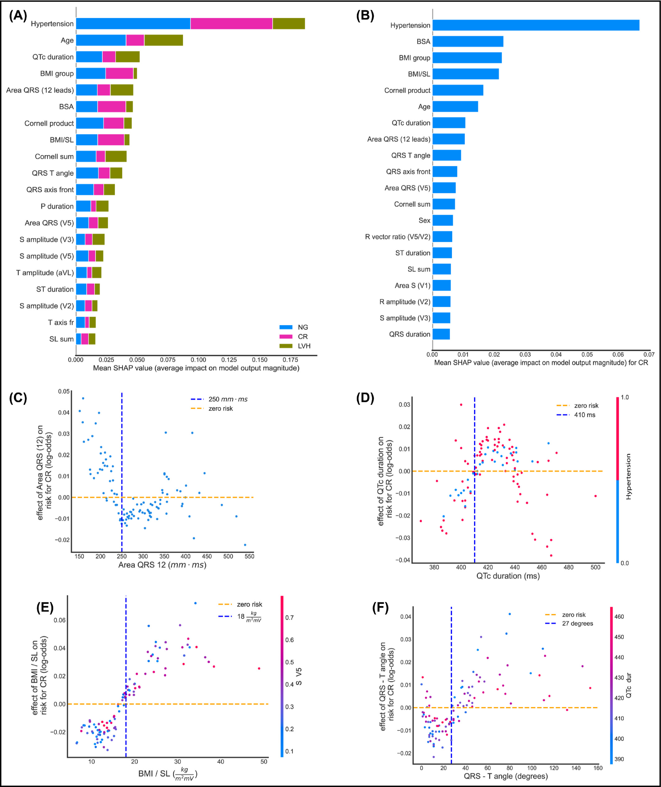
Cardiac remodeling is recognized as an important aspect of cardiovascular disease (CVD) progression. Machine learning (ML) techniques were applied to basic clinical parameters and electrocardiographic features, in order to detect abnormal left ventricular geometry (LVG) even before the onset of left ventricular hypertrophy (LVH), in a population without established CVD. The authors enrolled 528 patients with and without essential hypertension, but no other indications of CVD. All patients underwent a full echocardiographic evaluation and were classified into 3 groups; normal geometry (NG), concentric remodeling without LVH (CR), and LVH. Abnormal LVG was identified as increased relative wall thickness (RWT) and/or left ventricular mass index (LVMi). The authors trained supervised ML models to classify patients with abnormal LVG and calculated SHAP values to perform feature importance and interaction analysis. Hypertension, age, body mass index over the Sokolow‐Lyon voltage, QRS‐T angle, and QTc duration were some of the most important features. Our model was able to distinguish NG from CR+LVH combined, with 87% accuracy on an unseen test set, 75% specificity, 97% sensitivity, and area under the receiver operating curve (AUC/ROC) equal to 0.91. The authors also trained our model to classify NG and CR (NG + CR) against those with LVH, with 89% test set accuracy, 93% specificity, 67% sensitivity, and an AUC/ROC value of 0.89, for a 0.4 decision threshold. Our ML algorithm effectively detects abnormal LVG even at early stages. Innovative solutions are needed to improve risk stratification of patients without established CVD, and ML may enable progress in this direction.

